Ultrasound In Pregnancy
Ultrasound is one of the most widely used tests to view images and monitor embryonic development during pregnancy. This technique is harmless for the fetus and allows early detection of possible alterations.
Other imaging tests are contraindicated in pregnancy, as they can cause abnormalities in the fetus. These tests are radiographs that use X-rays and computerized axial tomography (CT), which is based on the use of ionizing radiation.
The first ultrasound is usually done at the first gynecological consultation to confirm pregnancy, since it allows us to see the gestational sac where the embryo will develop.
Once the pregnancy is confirmed, an ultrasound is scheduled for each trimester, except in high-risk pregnancies.
Let’s see more below.
What is an ultrasound?
It is a diagnostic technique that is based on the use of ultrasound.
Ultrasounds are waves with a wavelength shorter than that perceived by the human ear. They are mechanical waves, that is, non-ionizing, which means that they are not harmful to body tissues.
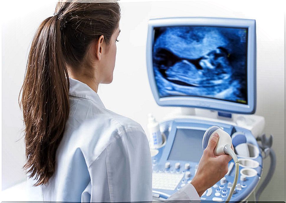
Ultrasound machines consist of a transducer that emits ultrasound and receives the echoes they produce. These echoes are transformed into electrical energy that is later represented in an image on the screen.
Current devices also allow selecting the intensity of ultrasound, adapting the characteristics of the wave to the organ we want to explore.
The transducer is placed in the area to be scanned and a representation of it is seen on the screen.
Moving the transducer, the area to be tested is searched for. The image that appears on the screen allows us to see the structure we are studying in real time, so it has multiple applications.
First trimester ultrasound

It is carried out between 11 and 14 weeks of gestation and its objectives are as follows:
- Identify the number of embryos and, in the case of multiple gestation, see if they are univitelline (twins) or polyviteline (twins).
- Identify the heartbeat, checking the viability of the pregnancy. The embryonic heart beats from 22 days of gestation, perceiving this beat with the ultrasound allows to confirm that the embryo is alive.
- Visualize embryonic movements, which appear from the ninth week of development and contribute to confirm the vitality of the embryo.
- Estimate the gestational age.
Although the date of the last menstruation orients on the age of the embryo, the precise age is made with ultrasound parameters, such as the measurement of the cranio-caudal axis. In this parameter it measures the distance between the head and the buttocks of the embryo.
- Rule out chromosomal malformations.
The first trimester ultrasound is part of the well-known triple screening, which allows early suspicion of the existence of chromosomal diseases such as Down syndrome.
- Observe the morphology of the embryo, checking that the development so far has not suffered alterations.
- See possible abnormalities of the uterus that could interfere with pregnancy, such as fibroids.
Second trimester ultrasound

This ultrasound is done between 16 and 20 weeks of pregnancy. In this case it is usually abdominal.
At this stage, the external morphology of the embryo is practically developed, so that structural alterations can be detected. For this reason, this ultrasound is also called a morphological ultrasound.
If any possible structural alteration is found, other tests are carried out to confirm or guide it towards the cause.
Also, if the position of the fetus allows it, it may be possible to know its sex.
Third trimester ultrasound

It is the last ultrasound control of pregnancy and is performed between weeks 32 and 36 of gestation.
During the last weeks of embryonic development there should be significant growth of the fetus, so this ultrasound seeks to calculate the weight and fetal growth, checking that it is adequate.
Regarding delivery, it is interesting to see the fetal statics, that is, what position the fetus has within the uterus. The most common is that the fetus is in a cephalic position, with the head directed towards the cervix.
Sometimes the fetus is transverse or breech, which may make a cesarean section recommended.
In this ultrasound the mother’s uterus is also inspected, ruling out pathologies that may complicate the end of pregnancy, such as a placental abruption or a placenta previa.
Types of ultrasound
- Transabdominal: it is done abdominally, placing the transducer on the lower part of the abdomen.
- Transvaginal: the transducer is inserted through the vagina.
In special cases, another type of ultrasound is used:
- Doppler ultrasound: usually used in the last trimester if you want to monitor blood flow through the umbilical cord.
- 3D Ultrasound – This three-dimensional ultrasound produces thousands of near-simultaneous photos, which together produce a near-photographic image.
- 4D ultrasound: It is similar to 3D, but produces the image on video, to allow you to see the movements of the fetus.
In short, ultrasound is a recommended test for all pregnant women, since it allows knowing interesting and useful data about their health and, of course, the fetus.